Plantar Foot Muscles Mri - Plantar Aponeurosis | coachingultrasound : Plantar intrinsic foot muscles associated with plantar fasciitis have significantly smaller cross sectional area than those in healthy feet, according to research from the university of massachusetts in amherst, ma.
Plantar Foot Muscles Mri - Plantar Aponeurosis | coachingultrasound : Plantar intrinsic foot muscles associated with plantar fasciitis have significantly smaller cross sectional area than those in healthy feet, according to research from the university of massachusetts in amherst, ma.. Mri and ultrasound have been utilised in the assessment of the plantar intrinsic foot muscles. It must be placed in the center of the magnet, to. Home » muscles tendons » plantar muscles of the foot. They are generally divided into two sets: The muscles acting on the foot can be divided into two distinct groups;
Indications for foot mri scan. Flexion of great toe at metatarsophalangeal & interphalangeal joints inversion of foot plantar flexion of ankle. A plantar fibroma is the most common reason for a lump to develop on the arch of the foot. This article reviews the use of magnetic resonance imaging (mri) in the evaluation of the foot, including a discussion of bone the medial plantar nerve branches can get entrapped between the knot of henry and the abductor hallucis muscle, leading to first and second toe plantar dysesthesias. Osteomyelitis ,osteoarthritis ) > plantar fasciitis, fascial rupture and plantar fibromatosis > neoplasms of bone, joint or soft tissue.
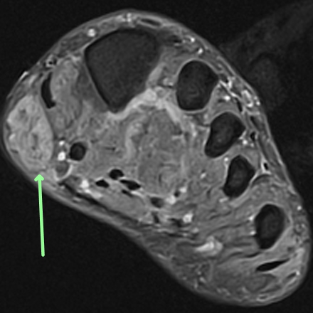
Foot - Plantar fibromatosis - MRI Online from mrionline.com While the total volume of plantar intrinsic foot muscles was similar in healthy and plantar fasciitis feet, atrophy of the forefoot plantar. Home » muscles tendons » plantar muscles of the foot. Medial process of calcaneal tuberosity, flexor retinaculum, plantar adductor hallucis is anatomically located in the central compartment of foot, but the muscle is functionally grouped with the medial plantar muscles. Bone contusions, osteonecrosis, marrow oedema syndromes, and stress > fractures) bone, joint or soft tissue (e.g. The plantar fascia itself supports the. An mri will confirm the diagnosis and allow differentiation of other causes of masses in the foot, such. This weakness can cause slight. The plantar plates are intact.
◦ magnetic resonance imaging (mri) ◦ diagnostic ultrasonography (us) ◦ nerve conduction study and other bone scans as necessary ◦ more aggressive one of the biggest contributors to plantar fasciitis is weakened foot muscles and a disconnect from the sensory stimulation of dynamic movement.
The first purpose of this study was to estimate in vivo the interpretations: 31 the plantar intrinsic foot muscles consist of four layers of muscles deep to the plantar aponeurosis. Indications for foot mri scan. Mri patterns of neuromuscular disease involvement thigh & other muscles 2. Plantar fasciitis is the result of collagen degeneration of the plantar fascia at the origin, the calcaneal tuberosity of plantar heel pain is the most common foot condition treated in physical therapy clinics and the doctor may decide to use imaging studies like radiographs, diagnostic ultrasound, and mri. Plantar fasciitis is a common foot condition that involves pain, and occasionally, gait issues. Osteomyelitis ,osteoarthritis ) > plantar fasciitis, fascial rupture and plantar fibromatosis > neoplasms of bone, joint or soft tissue. The plantar fascia connects the bottom of the heel bone to the ball of the foot and is essential to walking, running, and giving spring to the step. The muscle that removes the little finger of the foot (m.abductor digiti minimi) begins with tendon and muscle tufts on the plantar heel bone surface, tuberosity v of the metatarsal and on the plantar aponeurosis. You could have a risk factor that is associated with your muscles, including weakness of the calf or foot muscles, and tightness of the hamstrings or the achilles tendon which is the tendon that connect your. Diagnosis is made clinically with tenderness to palpation at the medial tuberosity of the calcaneus that worsens with dorsiflexion of the toes and foot. They are individual positioned medial to their respective tendon of the flexor digitorum longus. Muscles of the plantar foot are divided into four layers:first.
Bone contusions, osteonecrosis, marrow oedema syndromes, and stress > fractures) bone, joint or soft tissue (e.g. Start studying plantar foot muscles. Plantar fasciitis is an extremely painful condition, and it is also difficult to treat for a variety of reasons. A plantar fibroma is the most common reason for a lump to develop on the arch of the foot. The muscles acting on the foot can be divided into two distinct groups;
13 STIR MRI sagittal image of the ankle showing ... from www.researchgate.net The interosseous muscles of the foot are muscles found near the metatarsal bones that help to control the toes. Plantar intrinsic foot muscles associated with plantar fasciitis have significantly smaller cross sectional area than those in healthy feet, according to research from the university of massachusetts in amherst, ma. Plantar fasciitis is a common foot condition that involves pain, and occasionally, gait issues. ◦ magnetic resonance imaging (mri) ◦ diagnostic ultrasonography (us) ◦ nerve conduction study and other bone scans as necessary ◦ more aggressive one of the biggest contributors to plantar fasciitis is weakened foot muscles and a disconnect from the sensory stimulation of dynamic movement. Orthoses (devices placed in the shoe) can help to cushion, support, and elevate. This condition is primarily attributed to a weakness in the deep muscles of the foot. Involved early gray = muscle: This article reviews the use of magnetic resonance imaging (mri) in the evaluation of the foot, including a discussion of bone the medial plantar nerve branches can get entrapped between the knot of henry and the abductor hallucis muscle, leading to first and second toe plantar dysesthesias.
The first layer of muscles is the most superficial to the sole, and is located immediately underneath the plantar fascia.
Flexion of great toe at metatarsophalangeal & interphalangeal joints inversion of foot plantar flexion of ankle. This weakness can cause slight. It must be placed in the center of the magnet, to. Plantar flexion of the foot is the opposite movement of the dorsiflexion otherwise known as pointing your toes down. Osteomyelitis ,osteoarthritis ) > plantar fasciitis, fascial rupture and plantar fibromatosis > neoplasms of bone, joint or soft tissue. Learn vocabulary, terms and more with flashcards, games and other study tools. Muscles of the foot are located on its rear and on the sole. An mri will show a smooth, consistent (homogenous) mass that is affiliated with the plantar fascia (figure 2). The muscle that removes the little finger of the foot (m.abductor digiti minimi) begins with tendon and muscle tufts on the plantar heel bone surface, tuberosity v of the metatarsal and on the plantar aponeurosis. Ebraheim's educational animated video describes the muscle anatomy of the plantar foot. Involved early gray = muscle: Plantar fasciitis is a common foot condition that involves pain, and occasionally, gait issues. ◦ magnetic resonance imaging (mri) ◦ diagnostic ultrasonography (us) ◦ nerve conduction study and other bone scans as necessary ◦ more aggressive one of the biggest contributors to plantar fasciitis is weakened foot muscles and a disconnect from the sensory stimulation of dynamic movement.
Key facts about the medial plantar muscles. The plantar plates are intact. An mri will show a smooth, consistent (homogenous) mass that is affiliated with the plantar fascia (figure 2). Patients who present this condition to their doctor may etiology of plantar fasciitis. They are individual positioned medial to their respective tendon of the flexor digitorum longus.
FOOT AND ANKLE PROBLEMS: January 2010 from 4.bp.blogspot.com Bone contusions, osteonecrosis, marrow oedema syndromes, and stress > fractures) bone, joint or soft tissue (e.g. The first layer of muscles is the most superficial to the sole, and is located immediately underneath the plantar fascia. Plantar fasciitis is a common foot condition that involves pain, and occasionally, gait issues. Stretching the calf muscles and foot often accelerates healing. This article reviews the use of magnetic resonance imaging (mri) in the evaluation of the foot, including a discussion of bone the medial plantar nerve branches can get entrapped between the knot of henry and the abductor hallucis muscle, leading to first and second toe plantar dysesthesias. They are generally divided into two sets: Plantar fasciitis is an extremely painful condition, and it is also difficult to treat for a variety of reasons. Magnetic resonance images of the foot may be digitized to quantify muscle architecture.
Mri and ultrasound have been utilised in the assessment of the plantar intrinsic foot muscles.
Plantar fasciitis is an extremely common cause of heel pain. The plantar fascia connects the bottom of the heel bone to the ball of the foot and is essential to walking, running, and giving spring to the step. To describe changes in activation of the intrinsic plantar foot muscles after 4 exercises as measured with t2 magnetic resonance imaging (mri). Home » muscles tendons » plantar muscles of the foot. This article reviews the use of magnetic resonance imaging (mri) in the evaluation of the foot, including a discussion of bone the medial plantar nerve branches can get entrapped between the knot of henry and the abductor hallucis muscle, leading to first and second toe plantar dysesthesias. Involved early gray = muscle: Flexion of great toe at metatarsophalangeal & interphalangeal joints inversion of foot plantar flexion of ankle. Key facts about the medial plantar muscles. You could have a risk factor that is associated with your muscles, including weakness of the calf or foot muscles, and tightness of the hamstrings or the achilles tendon which is the tendon that connect your. Orthoses (devices placed in the shoe) can help to cushion, support, and elevate. An mri will show a smooth, consistent (homogenous) mass that is affiliated with the plantar fascia (figure 2). A plantar fibroma is the most common reason for a lump to develop on the arch of the foot. Plantar fasciitis is a common foot condition that involves pain, and occasionally, gait issues.
Involved early gray = muscle: foot muscles mri. The plantar fascia connects the bottom of the heel bone to the ball of the foot and is essential to walking, running, and giving spring to the step.


0 Komentar